DIJITAL RONTGEN GE OPTİMA XR646
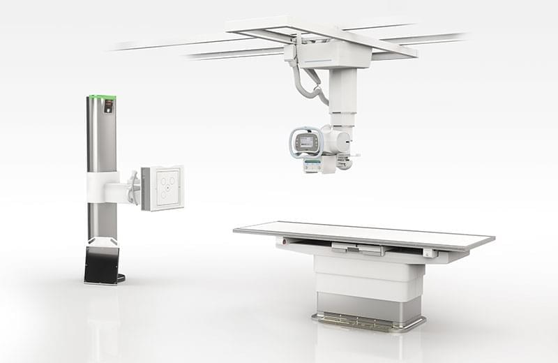
It is a top-of-the-line digital radiography system with a wireless Flashpad detector that enables the daily challenges of imaging to be overcome efficiently. This system, which stands out from the others with its most up-to-date optimization technologies, allows quick and flexible positioning of patients and devices, allowing rapid and safe initiation of examinations. The system, which can easily and effortlessly perform the editing of images and transferring the examinations, protects the comfort of the patients and ensures that they are always under care.
The Optima XR646 has been designed to facilitate and shorten the work of the technicians in all inspection steps and to achieve more satisfactory results. Thanks to its simplified examination features and motorized vertical movement capabilities, Flashpad allows quick and easy positioning of patients with the wireless detector. You can preview the image in less than 3 seconds to instantly evaluate the image quality and avoid the need for retakes. Work more efficiently, reduce bottlenecks and shorten patients' waiting times thanks to the system that gives the whole image in 10 seconds. During reviews, you can switch back and forth between the workstation keyboard and the touchscreen interface. For patients who want to see their entire examination on a single page, you can print multiple images on a single page, and you can ensure that the critical distance between source and image is precisely maintained regardless of the patient's position. Thanks to automatic tracking, when correction is required in positioning, the tube and detector are automatically aligned to ensure correct source-image distance. In this way, the possibility of re-shooting is reduced and your workflow is accelerated. Thanks to the real Dual Energy feature, which performs two shots in one shot with 200 ms intervals in the system, in addition to the standard radiography image, soft tissue images free of bone tissue and an image in which bone or foreign body or calcified objects can be seen can be obtained. In addition, thanks to the fully automatic scoliosis / long bone extraction feature, subsequent images can be taken and combined, so a single image can be created to evaluate the relevant anatomy in orthopedic examinations.
MOBILE X-RAY GE OPTIMA XR240amx
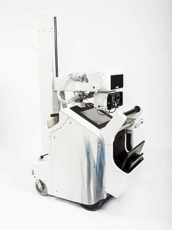
The Optima XR240, a motorized mobile x-ray system with ergonomic design, smart power management and wireless connectivity providing seamless workflow, works with the high image quality Flashpad HD digital wireless detector from GE Healthcare and is a product of the over 18,000 AMX family that are still in use. While developing the AMX Optima XR240 system, to take our systems further; We asked technicians, doctors and managers some questions and observed work habits. Thanks to this research, we discovered methods to help you do your job more easily and efficiently. As a result; We have seen the demand for a system that operates through common sense and offers high image quality. Based on this, we developed the AMX Optima XR240, which offers more advanced X-rays. Small footprint, flexible movement and offering optimized workflows in a 24/7 mobile environment are the main features of this system.
M530 OHX Premium Operating Microscope
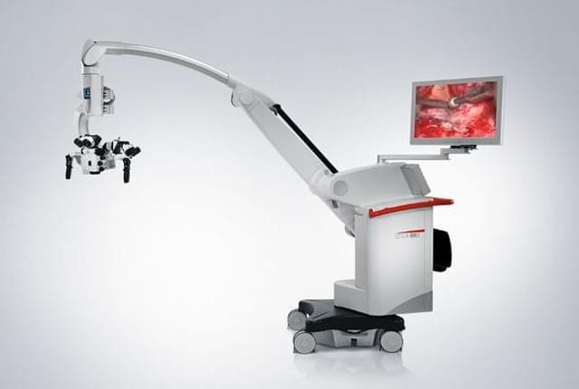
- Fluorescence module for neuro-oncological use guided by 5-ALA active ingredient
- Fluorescence module for neuro-vascular and neuro-oncological use guided by Sodium Fluorescein
- Fluorescence module for ICG-guided neuro-vascular use
- Wireless foot pedal
PENTAX IMAGINA ENDOSCOPY
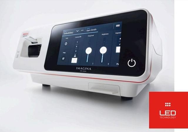
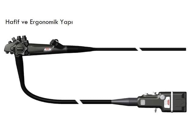
- High maneuverability
- Areal enhanced imaging modes (rectum, duodenum, colon, gastro esophagus, vascular)
- The only system that does not use fiberoptic cables using LED technology at the scope tip
- 180º image rotation
- 3 different texture inspection modes (Tone Enhancement, Surface Enhancement, Contrast Enhancement)
- Simultaneous viewing of the live image and the recorded video image (It can be viewed on the digital screen of the device or on the monitor)
BT GE Revolution CT
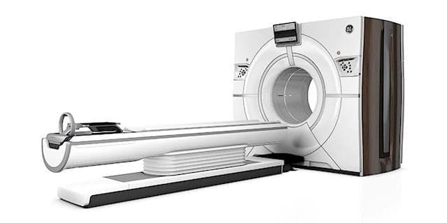
The 512-Fractional BT system serving in our hospital offers our patients the most up-to-date technology that allows the use of "Computer Aided Detection (CAD)" software. In this way, personalized image reconstructions can be obtained as dedicated to pathology.
In addition to having the largest imaging detector used in Computed Tomography systems, Revolution CT is a unique technology that uses different bit detector materials for the first time since the invention of CT technology. A pioneering design was used in the system, which is also referred to as "Diamond Detector". In addition, it is equipped with a special collimator technology that minimizes the radiation scattered during shooting, thanks to the 3D collimator developed for the patient to receive less radiation.
Revolution CT can collect 101 separate energy level data from the patient in one rotation, using a method called Ultra Fast kV Switching technology. In this way, unlike standard IT technologies, it also provides information about the structure of the tissue.
Revolution CT performs cardiovascular angiography in a single beat not only in low pulse beats but also in high pulses. In this way, not only adults but also pediatric patient group cardiac angiographies can be performed under CT.
images of high quality and without image distortion (artifact-free) can now be obtained even in patients with prosthetics or implants. Even in our patients with hip prostheses, other tissues around the implant can be monitored in high quality without image distortion. In this way, it is very easy to perform the necessary controls and the location of the nails in patients who have undergone waist nailing.
Revolution CT has the ability to dose individually according to each patient's own body structure. In this way, the adult / pediatric patient group is displayed with doses suitable for body size and shape. In single shots where more than one organ is scanned, it receives rays according to the density of each organ of the body. In this way, patient safety is ensured at the maximum level.
Revolution CT has shooting protocols specially planned for adults and ages. Revolution CT user has the opportunity to work with appropriate acquisition parameters according to the age and weight of the patient. In this way, patient safety and health is placed first.
Revolution CT not only with its superior technical features, but also allows the patient to complete his / her experience from beginning to end with maximum comfort. The tunnel width of Revolution CT is 80 cm. In this way, even patients with fear of closed spaces can be performed very easily.
Revolution CT allows the use of much less contrast agent in patient groups at risk of kidney failure. With the Gemstone Spectral Imaging method, the system recognizes the contrast material performed from the patient's vein and reconstructs the images as if much more contrast material was used. In this way, the use of contrast agents in both adult cases with a risk of kidney failure and in pediatric cases is controlled.
These are the successful applications of Revolution CT, with the protection of X-ray sensitive organs such as eyes, thyroid and breast. Eye, thyroid and breast tissue entering the image areas during the shooting of other organs are protected by ODM (Organ Dose Modulation) technology. The system automatically downloads the X-ray application during the shooting moments corresponding to these areas. In this way, sensitive organs are automatically protected by the system at a high level.
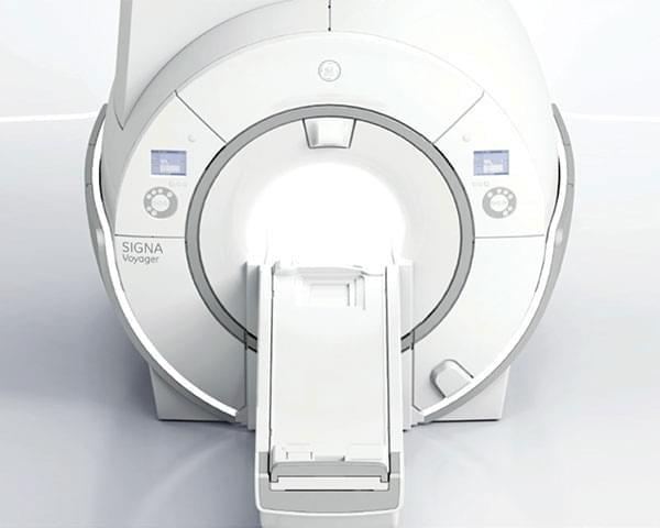
GE Healthcare 1.5 Tesla Voyager Magnetic Resonance Imaging System
The 1.5 Tesla General Electric Voyager G2 MR system started to be produced in 2018. Thanks to the 70 cm gantry opening, wide and spacious patient tunnel and the positioning of the patient's head outside the tunnel in most of the shots, it provides a significant reduction in claustrophobia cases. With TDI technology, effective signal values can be obtained with low noise rates with radio frequency reading algorithms.While receiving signals from antennas simultaneously with DST method, 16-channel new generation flex antennas can be filmed easily and quickly. It is a cause of serious discomfort in some patient groups. Adult and pediatric patients in this group can be viewed much more comfortably with silent MR software. In addition, moving tissues can be scanned with a special software without being affected by patient movements.
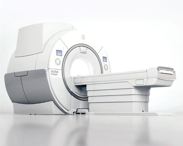
- This effect, which is called SAR- (Signal Absorbation Rate) and causes warming on the skin surface, is eliminated by the smart RF (Radio Frequency) management system used in Voyager systems. Thus, it enables both safe and more patient shooting per unit time.
- TDI Radio Frequency Technology – With the replacement of copper cables by fiber optic cables, MR devices started to produce faster and sharper results. Thanks to TDI technology, faster and more information can be collected from the patient in the same time compared to the past. Compared to old technology devices, the duration of the patient's stay in the MRI is reduced and it offers better quality imaging options.
- With the DST-Digital Surrounding Technology system, it is possible to display higher resolution in a shorter time with this solution, which is defined as the simultaneous operation of the signals received from both the body coil and the antennas embedded in the table.
- High Channel Flexible Coils – Thanks to the high channel and density light coils that adapt precisely to the patient's anatomy, even the most difficult shots are not impossible. Thanks to this coil technology, which replaces the old coils with low channels with heavy and positioning difficulties, patients can have a more comfortable MRI experience. - With the new generation coil apparatus developed for areas such as Shoulder, Knee, Ankle, Wrist, Elbow, it can be displayed with a much higher MR signal and higher resolution can be done in a shorter time.
- With the Multi Drive RF Technology - Application, as a result of the Radio Frequency signal sent from multiple sources stimulating the tissues gradually, overheating of the skin surface is prevented and signals can be received from deeper tissues.
- Silent Scan – Revolutionary silent MRI technology that reduces noise more than ever before, up to 3 decibels above ambient noise. It continues to create meaningful images for the clinician while providing the patient with a silent MRI experience with the help of high-channel and density coils. It is especially important for claustrophobic patients to overcome the fear of MRI and to reduce the amount of anesthesia used in pediatric patients.
- Advanced Motion Correction Techniques - Propeller MB provides motion regulation in all anatomic shots and reduces the possibility of repeating the shooting. Even if the patient moves during the shooting, the image is tried to be revealed without patient movements. This feature, which can also be used in dynamic imaging, reduces the patient's stay in MRI and provides a different MR experience for the patient.
- Imaging Around Implants – MAVRIC-SL enables advanced imaging of bones and soft tissues near implants.
- Focused Diffusion Weighted Imaging – FOCUS allows the use of small FOV sizes by using 2D selective stimulation pulses in shooting to avoid blurry images and low resolution problems caused by the use of wide FOV in diffusion imaging techniques, increasing the visibility of anatomical details with closer zoom and higher resolution. It is a new generation diffusion weighted imaging sequence. This sequence, which allows sharper results from the patient, eliminates the need for an endorectal coil in prostate imaging and provides a more tolerable MR experience.
- Liver Fatness Measurement - Ideal IQ is an advanced imaging sequence that can measure the percentage of triglyceride fat in the liver (Fat Fraction) with a percentage value, while simultaneously predicting measurement errors due to iron accumulation, thanks to T2 * correction.
- With Inhance MRI Angiography - applications, the brain, neck, abdomen and lower extremity (leg and thigh) parts can be visualized without medication.
- With the application called SWAN-, the distinction of cerebral hemorrhage and hemorrhage calcification can be demonstrated easily and without any medicated application.
- With the Navigator Body application, - the intraabdominal organs are monitored without being affected by patient movement, bowel movements and breathing movements, and provides a comfortable MRI experience especially to elderly patients, who are unconscious or unable to breathe.
- It is possible to view an area up to 180 cm with all Body-MRI sequences.
- With the new generation oil suppression technology called Ideal-4 different contrasts are created at the same time and oil printed shots can be made without any problem. This application increases the diagnostic reliability.
SIEMENS BIOGRAPH mCT
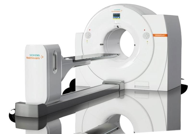
Biograph mCT whole body PET / CT system is designed for oncological imaging and diagnosis purposes. With its non-invasive imaging procedure, it provides detailed anatomical and physiological information at the molecular level as a result of the combined evaluation of PET and CT images. The system has 78 cm. Its gantry width allows PET scans to be performed more comfortably on patients. The multi-LSO detector structure, which provides 3D shooting, enables PET shooting with high sensitivity, high resolution, low radioactive dose and fast.
ELEKTA VERSA HD
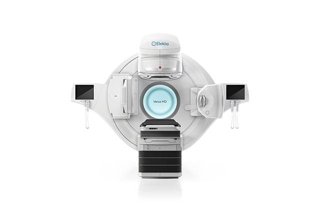
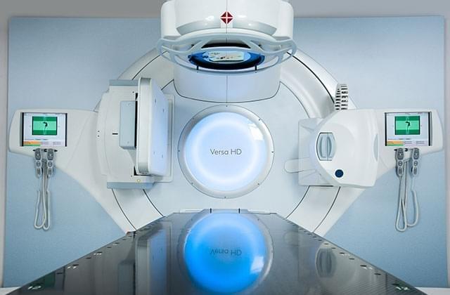
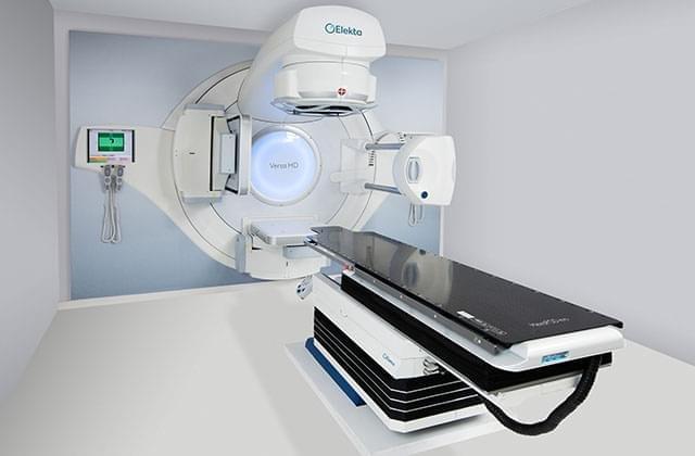
- With high definition dynamic radio surgery (HDRS), the Versa HD pushes your stereotactic capabilities.
- Performs stereotactic treatments with anatomically guided accuracy.
- Allows treatment with Versa HD, Monaco and high resolution MLC Agility in 40 x 40 cm area with 1 mm virtual leaves.
- With its methodology covering the whole process from start to finish, Versa HD allows even high doses to be delivered with high accuracy. Advanced 4D image guidance feature for lung and prostate SBRT